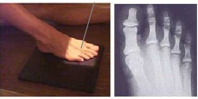
AP TOES PROJECTION
Anteroposterior • Dorsoplantar view • Specific evaluation of phalanges and interphalangeal joints
Exposure Factors
Very low exposure: Extremely low mAs due to minimal bone density of toes
Anatomical Structures Visible
Should be clearly observed:
- Distal phalanges - Terminal phalanges (nails)
- Middle phalanges - Intermediate phalanges (except hallux)
- Proximal phalanges - Basal phalanges
- Distal interphalangeal joints (DIP) - Between middle and distal phalanx
- Proximal interphalangeal joints (PIP) - Between proximal and middle phalanx
- Metatarsophalangeal joints (MP) - Base of toes
- Distal metatarsals - Heads of metatarsals
Cassette Size
Divided cassette: Transversely divided for multiple projections
Critical Angulation
Central ray must be angled cranially about 15° directed to the third metatarsophalangeal joint
This angulation is essential for optimal visualization of interphalangeal joint spaces
Patient Positioning
Central Ray Point
Direction: 15° cranial angulation
Location: Middle toe (third toe) MP joint
Goal: Optimal visualization of all phalanges and their joints
Toe Numbering and Phalanges
Toe 1 (Hallux)
Big toe - 2 phalanges
Toe 2
Second toe - 3 phalanges
Toe 3
Third toe - 3 phalanges
Toe 4
Fourth toe - 3 phalanges
Toe 5
Fifth toe - 3 phalanges
Patient Instructions
"You cannot move during the examination"
Keep toes completely still - Do not move toes during exposure
Optimal Image Characteristics
Expected superposition
Forearm over distal humerus
Visible structures
Distal humerus through superposition
Olecranon visible
Identifiable process
Adequate field
Complete joint included
Common Technical Challenges
Frequent problems in AP toes projection:
- Inadequate 15° angulation hiding interphalangeal joints
- Toe movement during exposure causing blurring
- Poor toe separation causing superposition
- Incorrect centering excluding important structures
- Overexposure due to extremely low bone density
- Foot rotation causing oblique view
Solution: Maintain precise 15° cranial angulation and ensure complete toe immobility
Special Technical Note
VERY LOW MILLIAMPERAGE (6 mAs)
Toes require extremely low mAs (6 mAs) due to:
- Minimal bone density - Very small and thin bones
- Practically no soft tissue - Very little X-ray attenuation
- High sensitivity to overexposure - Easy image burn
- Extreme fine detail required - Visualization of very small structures
- Low kV (42) - For maximum contrast in small structures